
Diagnosis of Fibroids
Ultrasound scan
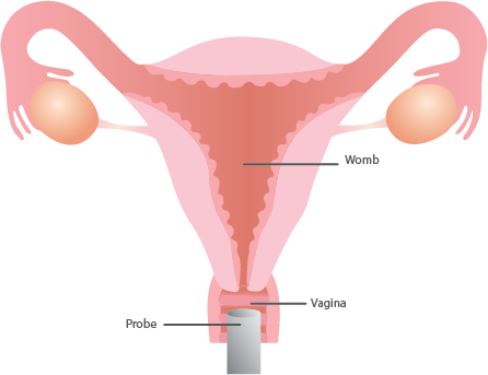
An ultrasound (sonography) of the pelvis can help in the diagnosis of Fibroids or other types of tumours. It can also provide more detailed information about the size and location of Fibroids.
This maybe an abdominal ultrasound, a vaginal ultrasound or both. An abdominal ultrasound is good at finding large Fibroids and for this you need to have a full bladder.. The scan itself is not painful as the probe is moved over the abdomen. A transvaginal ultrasound is usually the test used to diagnose Fibroids and here a narrow probe is put into the vagina .For a vaginal (internal) sonography there is no need for a full bladder.
Hysteroscopy
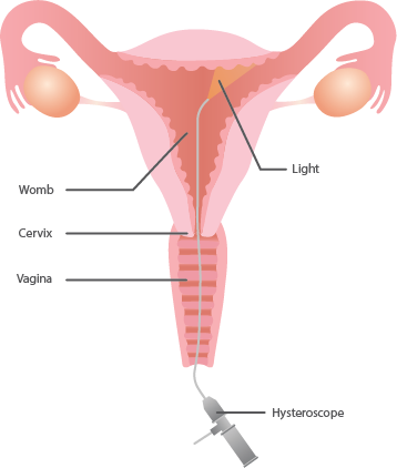
A hysteroscopy examines the inside of the womb (uterus) by using a small telescope (hysteroscope) which is inserted into your uterus through the vagina. Hysteroscopy can also be used to take a biopsy (tissue sample) of the lining of the womb. It can be done under local or general anaesthetic or in some cases, neither. Office hysteroscopy ,where a very thin hysteroscope is used can be done without any anaesthesia.. Hysteroscopy is done in hospital and the patient can usually go home the same day.
Laparoscopy
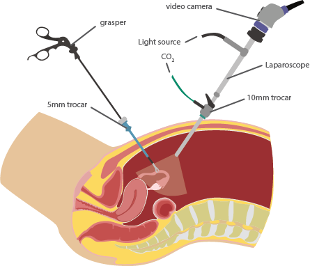
Laparoscopic surgery (‘Keyhole’ or ‘buttonhole’ surgery) can remove even large Fibroids with minimal cuts on the stomach wall. The entire surgery is done by watching the procedure on a television screen and operating with a camera in the abdomen. The large Fibroids are made into small pieces by means of a special machine called the morcellator and so can be removed through a small opening.
This procedure can be done in about 30-60 minutes under general anesthesia and small 2-3 cuts of ½ to 1 cm each. The patient’s hospitalization is less and recovery is very fast. Prolonged rest and absence from work is not needed.